Upper Thigh Muscles Ct Anatomy / Hip Muscle Strains Info Florida Orthopaedic Institute / Back thigh muscles of the gluteal and posterior femoral regions from gray's anatomy of the human body from 1918.
Upper Thigh Muscles Ct Anatomy / Hip Muscle Strains Info Florida Orthopaedic Institute / Back thigh muscles of the gluteal and posterior femoral regions from gray's anatomy of the human body from 1918.. There are different types of muscle, and some are controlled automatically by the. The uppermost of the medial thigh muscles is the pectineus muscle. Here we explain the major muscles of the human body. Case contributed by dr roberto schubert. Muscles are named according to their shape, location, or a combination.
Upper thigh muscles ct anatomy : The uppermost of the medial thigh muscles is the pectineus muscle. Muscular anatomy of upper limb dr bishnu khatiwada md resident radiodiagnosis, nams kathmandu, nepal 2. This article will introduce the muscles in each group and touch on their origin, insertion, function, and innervation. There are five muscles in the anterior thigh compartment:
Thigh Muscles Cross Sectional Anatomy Radiology Case Radiopaedia Org from prod-images-static.radiopaedia.org One further muscle of the anterior knee is the small articularis genus muscle, it is occasionally is blended with vastus intermedius. Sartorius, and the four quadriceps muscles; Iliopsoas psoas major psoas minor iliacus buttocks gluteal r. Upper thigh muscles ct anatomy / cross section through lower third of the upper arm. Introduction to functional anatomy of the hip flexors and anterior thigh muscles: Anatomy of the thigh and leg the thigh is best described in terms of compartmental anatomy, and is composed of anterior, posterior, and medial (adductor) compartments. Muscles in the medial thigh help to bring the thigh. The muscles of the lower limb are numerous and complex.
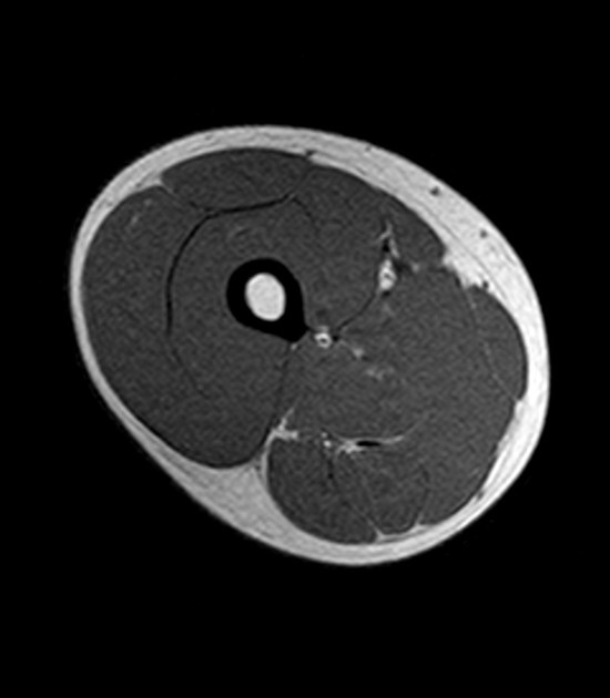
Case contributed by dr roberto schubert.
There are five muscles in the anterior thigh compartment: Here we explain the major muscles of the human body. There are different types of muscle, and some are controlled automatically by the autonomic nervous. The hamstring muscles in the back of the thigh, the quadriceps muscles in the front. The rectus femoris is located in the center of the thigh, while the vastus medialis is in the middle of the said body part. Each type of muscle tissue in the human body has a unique structure and a specific role. The four muscles all extend the lower leg. Simple and easy notes for quick revision. Other muscles of the anterior (front) thigh include the pectineus, sartorius, and the iliopsoas, which is made up of the psoas major and iliacus. This bone is very thick and. Muscles of the posterior cervical and upper thoracic spine 1. Introduction to functional anatomy of the upper extremity by joint action and exercise: On the anterior side, the most prominent of the muscles are the sartorius muscle and the four muscles that make up quadriceps muscle group (the quads.)
The uppermost of the medial thigh muscles is the pectineus muscle. 2, vastus medialis & intermedius muscles. Teachme anatomy part of the teachme series the medical information on this site is provided as an information resource only, and is not to be used or relied on for any diagnostic or treatment purposes. Muscles adapted for loaded versus unloaded actions. 2, vastus medialis & intermedius muscles.
1 from Muscles in the medial thigh help to bring the thigh. Muscular anatomy of upper limb dr bishnu khatiwada md resident radiodiagnosis, nams kathmandu, nepal 2. Simple and easy notes for quick revision. Proximal margin and deep surface of. Anatomically, it is part of the lower limb. The thigh is the area between the hip and the knee joint. 2, vastus medialis & intermedius muscles. The four muscles all extend the lower leg.
Muscular anatomy of upper limb dr bishnu khatiwada md resident radiodiagnosis, nams kathmandu, nepal 2.
Anatomically, it is part of the lower limb. Radiographers suggest an abdominal ct scan to look for the following: Proximal margin and deep surface of. This article will introduce the muscles in each group and touch on their origin, insertion, function, and innervation. Case contributed by dr roberto schubert. Upper thigh muscles ct anatomy / image result for iliacus muscle ct scan | ct scan : Anatomy of the thigh and leg the thigh is best described in terms of compartmental anatomy, and is composed of anterior, posterior, and medial (adductor) compartments. Written by keith bridwell, md; Upper thigh muscles ct anatomy : There are different types of muscle, and some are controlled automatically by the. The muscles that move the forearm are located along the humerus, which include the triceps brachii, biceps brachii, brachialis, and brachioradialis. .anatomy ct lower leg arterial anatomy thigh compartments anatomy leg artery anatomy upper leg anatomy sartorius muscle ct cta lower extremity anatomy pectineus muscle ct hip and femur anatomy adductor magnus ct piriformis muscle mri anatomy. Anatomical structures of the lower limb (hip, thigh, knee, leg, ankle and foot) and specific regions (compartment of the lower.
When a muscle is stretched beyond its limit, a tear can occur that can range from mild to serious. Related posts of muscle anatomy of upper thigh. 2, vastus medialis & intermedius muscles. Learn faster with these free muscle labeling diagrams. There are five muscles in the anterior thigh compartment:
Normal Mri Of The Thigh Radiology Case Radiopaedia Org from prod-images-static.radiopaedia.org Anatomical structures of the lower limb (hip, thigh, knee, leg, ankle and foot) and specific regions (compartment of the lower. The four muscles all extend the lower leg. The sartorius muscle can cause and contribute to burning stinging down the thigh to the inside of the knee. Muscles adapted for loaded versus unloaded actions. Lower limbs radiology key / almost all muscles cross at least one joint (moveable connection between two bones) and cause an action across that joint. Abdominal computed tomography (ct) is a type of medical imaging procedure used to diagnose and monitor internal stomach issues, like cancer, bowel obstruction, and abdominal pain. Proximal margin and deep surface of. Upper thigh muscles ct anatomy.
Ct, cartilage, and bone histology, joints, upper limb anatomy 1 (text only).
Muscular anatomy of upper limb dr bishnu khatiwada md resident radiodiagnosis, nams kathmandu, nepal 2. .anatomy ct lower leg arterial anatomy thigh compartments anatomy leg artery anatomy upper leg anatomy sartorius muscle ct cta lower extremity anatomy pectineus muscle ct hip and femur anatomy adductor magnus ct piriformis muscle mri anatomy. The muscles that move the forearm are located along the humerus, which include the triceps brachii, biceps brachii, brachialis. Simple and easy notes for quick revision. As the name implies they adduct the thigh at the hip. Anatomy of the thigh and leg the thigh is best described in terms of compartmental anatomy, and is composed of anterior, posterior, and medial (adductor) compartments. There are five muscles in the anterior thigh compartment: There are different types of muscle, and some are controlled automatically by the. It is part of the lower limb. This article will introduce the muscles in each group and touch on their origin, insertion, function, and innervation. On the anterior side, the most prominent of the muscles are the sartorius muscle and the four muscles that make up quadriceps muscle group (the quads.) 2, vastus medialis & intermedius muscles. The thigh is the area between the hip and the knee joint.
Lower limbs radiology key / almost all muscles cross at least one joint (moveable connection between two bones) and cause an action across that joint upper thigh anatomy. Related posts of muscle anatomy of upper thigh.


0 Komentar